
Understanding Your Heart and How it Functions Cardiology
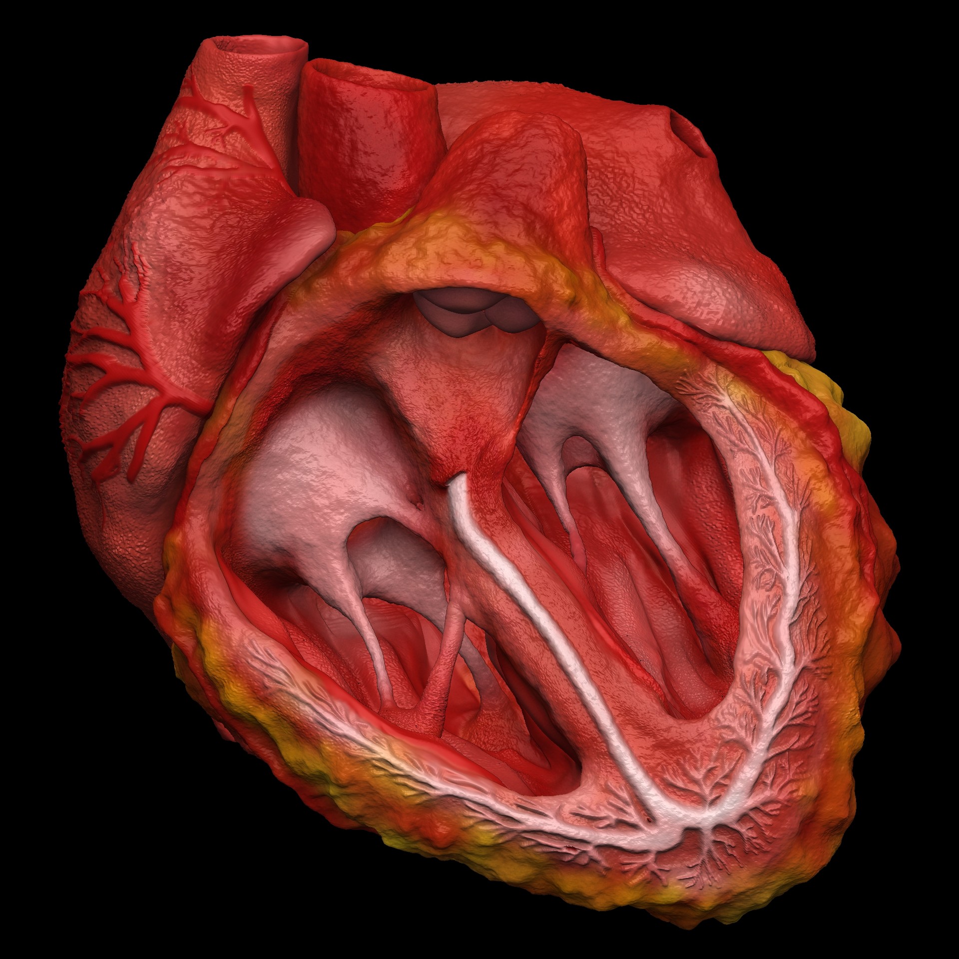
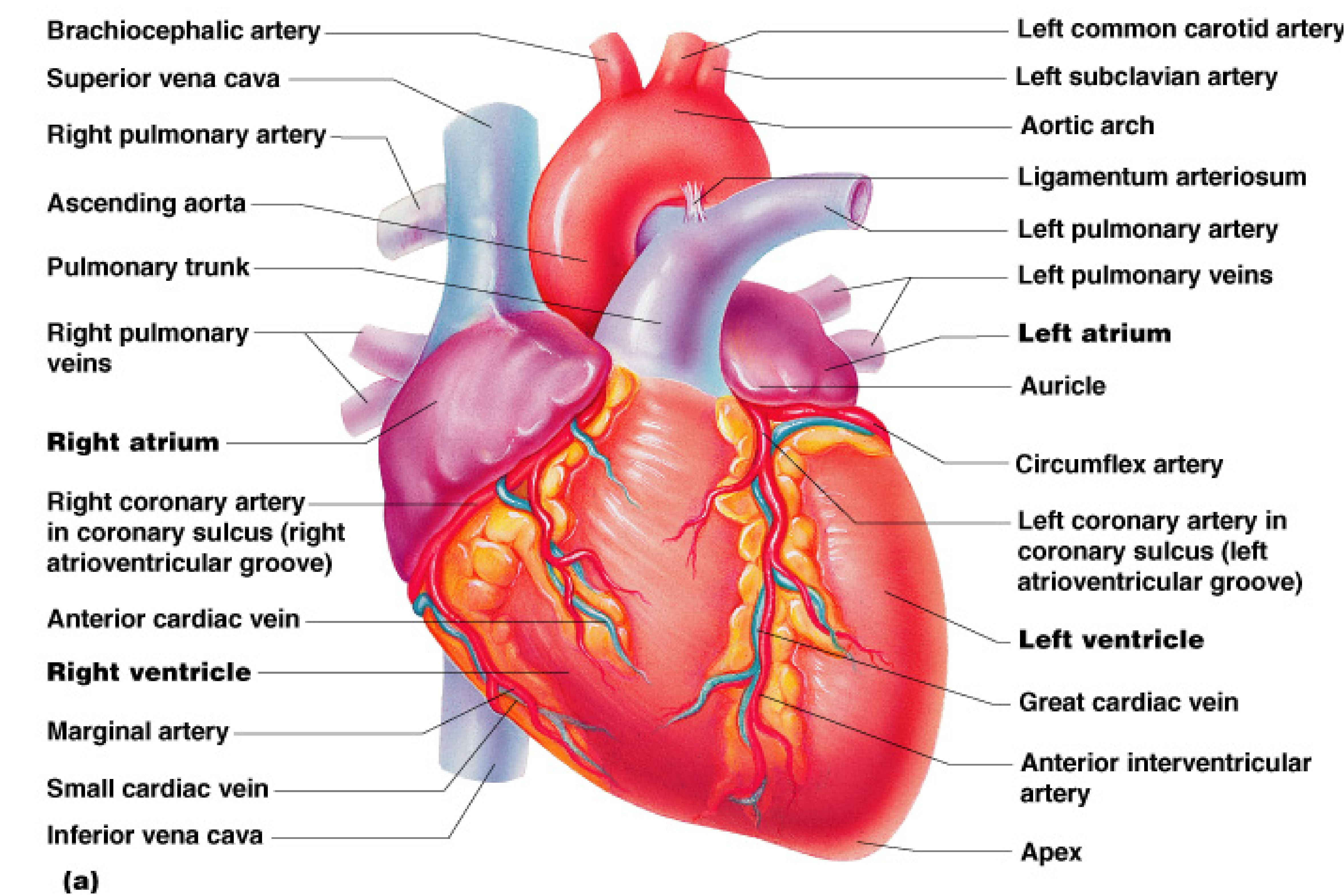
The cusps are pushed open to allow blood flow in one direction, and then closed to seal the orifices and prevent the backflow of blood. Backward prolapse of the cusps is prevented by the chordae tendineae-also known as the heart strings-fibrous cords that connect the papillary muscles of the ventricular wall to the atrioventricular valves.. There are two sets of valves: atrioventricular.

7 Beautiful Human Heart 3d Model Free Download Viela Mockup
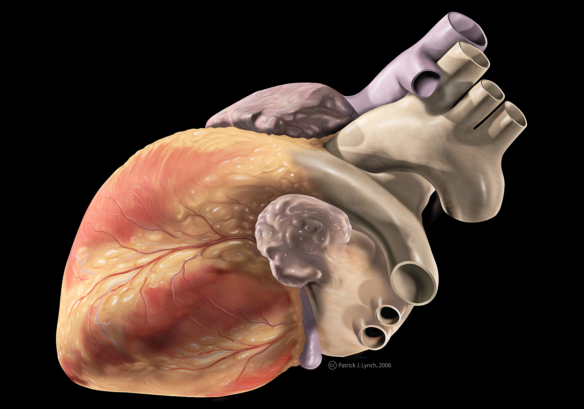
Real-time MRI of the human heart The human heart is in the middle of the thorax, with its apex pointing to the left. The human heart is situated in the mediastinum, at the level of thoracic vertebrae T5-T8. A double-membraned sac called the pericardium surrounds the heart and attaches to the mediastinum.

Human Heart 3D model circulatory CGTrader
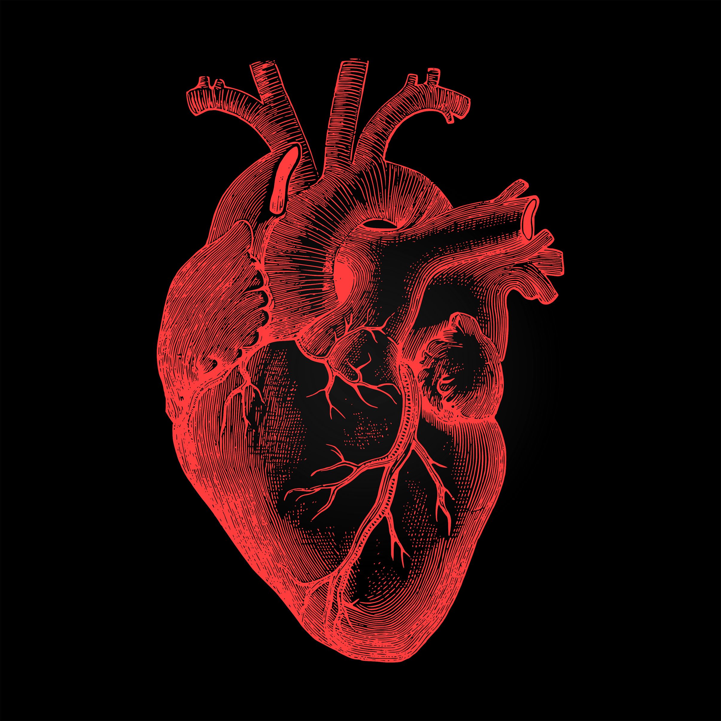
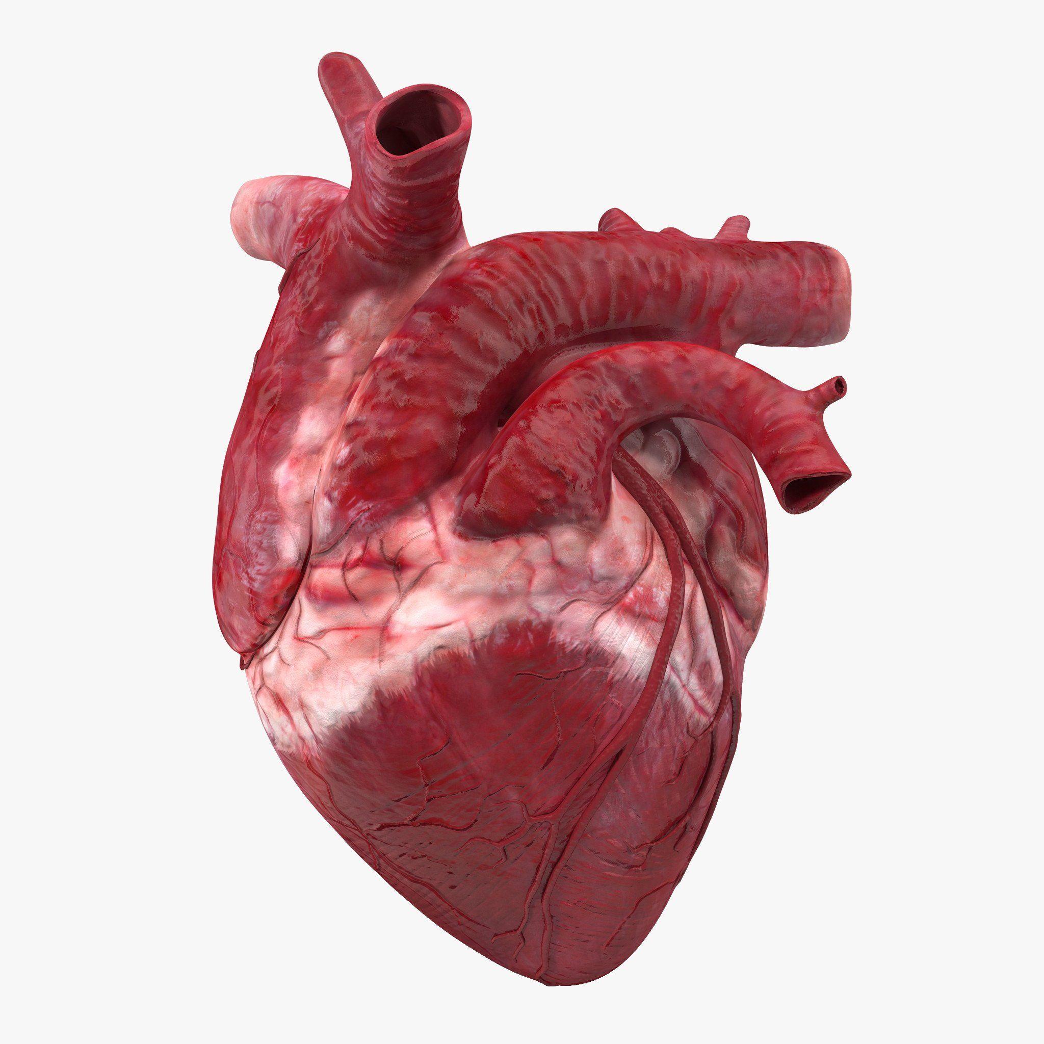
Heart beat. The human heart is a fist-sized muscle with a rounded bottom, smooth sides, and a thick arch of blood vessels at the top. So how did it come to be represented around the world by the pointy-bottomed, smooth-sided, cleft-topped icon drawn by doodlers, artists, and greeting card makers? No one really knows.
humanheartanatomicalrenderingondarkbackground TrendinTech
Why does the heart pump blood? Where is the heart actually located? Take a virtual look into the heart and get answers to all your questions. This video show.
Human Heart 3d Model on Behance
Browse Getty Images' premium collection of high-quality, authentic Human Heart stock photos, royalty-free images, and pictures. Human Heart stock photos are available in a variety of sizes and formats to fit your needs.
Human Heart Photograph by Medimation/science Photo Library Fine Art America
Your heart muscle is made up of three layers of tissue: Pericardium - a thin, outer lining that protects and surrounds your heart.; Myocardium - a thick, muscular middle layer that contracts and relaxes to pump blood around of your heart.; Endocardium - a thin, inner layer that makes up the lining of the four chambers and the valves in your heart..
Heart Contractions Simplified Interactive Biology, with Leslie Samuel
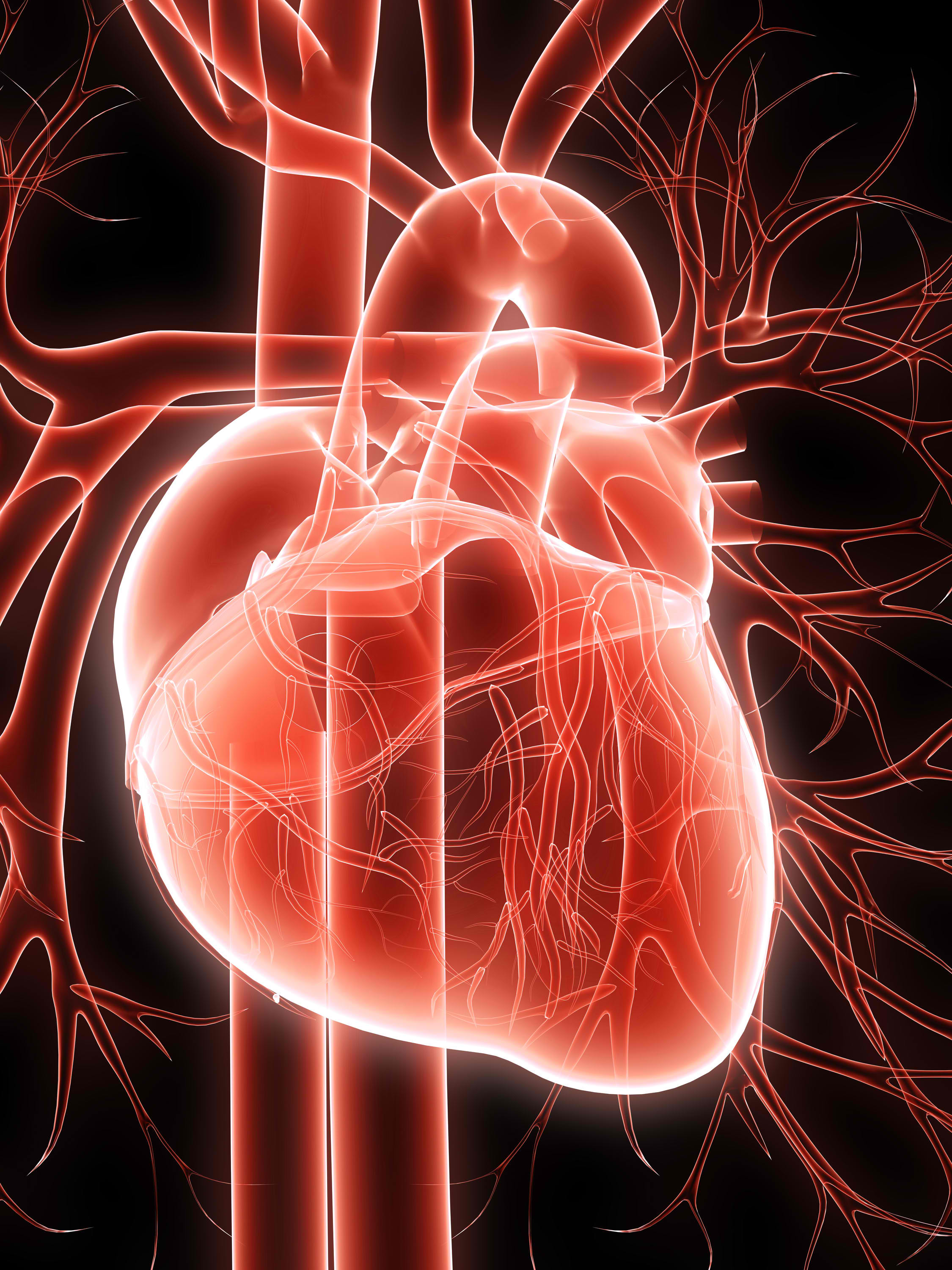
Your heart is the primary organ of your circulatory system. It pumps blood throughout your body, controls your heart rate and maintains blood pressure. Your heart is a bit like a house. It has walls, rooms, doors, plumbing and an electrical system. All the parts of your heart work together to keep blood flowing and send nutrients to your other.

Structure Of Human Heart Vector Illustration Of Diagram Of Human Heart Anatomy Heart is
The heart is made of three layers of tissue. Endocardium is the thin inner lining of the heart chambers and also forms the surface of the valves.; Myocardium is the thick middle layer of muscle that allows your heart chambers to contract and relax to pump blood to your body.; Pericardium is the sac that surrounds your heart. Made of thin layers of tissue, it holds the heart in place and.

Structure Of Human Heart Vector Illustration Of Diagram Of Human Heart Anatomy Heart is
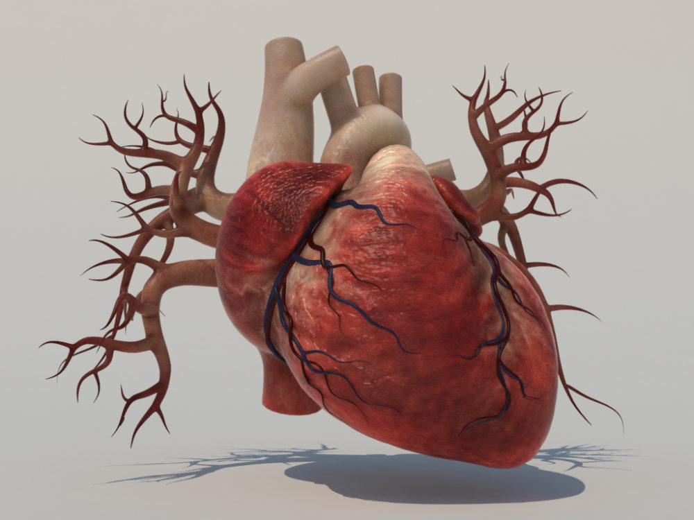
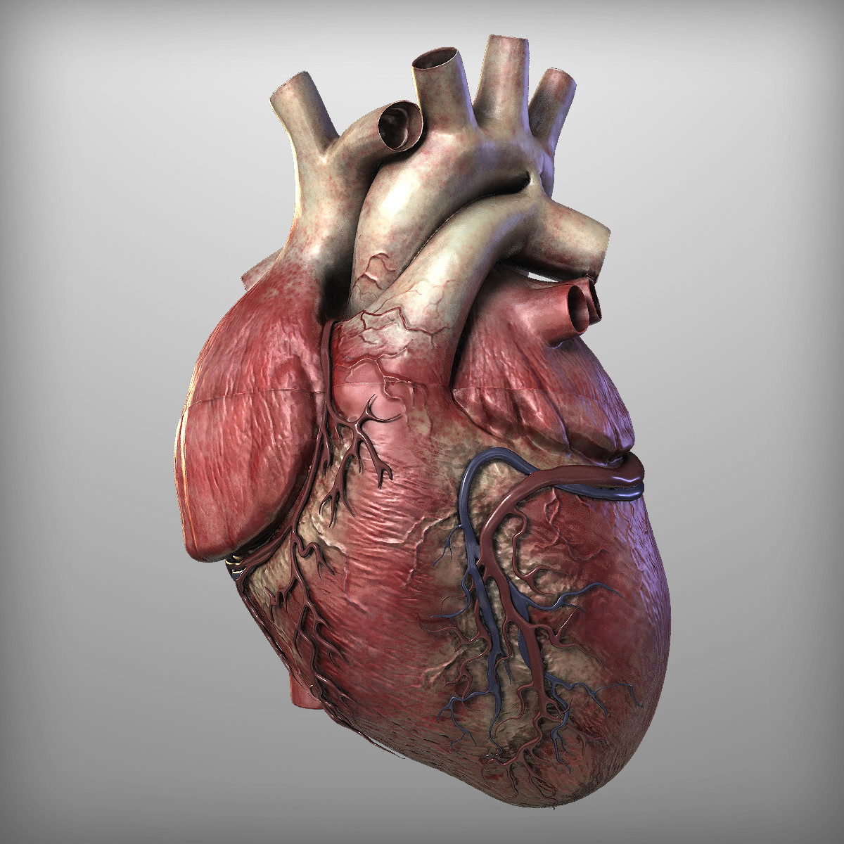
The heart is the body's engine room, responsible for pumping life-sustaining blood via a 60,000-mile-long (97,000-kilometer-long) network of vessels.
Heart Anatomy Wallpapers Wallpaper Cave
This makes the Carmat heart much more vulnerable compared to other artificial hearts. And as always, advantages come at a price. At around $200,000 (€183,000) per heart, the price is not exactly.
Human Heart Images Real imgextra
Browse Getty Images' premium collection of high-quality, authentic Real Human Heart stock photos, royalty-free images, and pictures. Real Human Heart stock photos are available in a variety of sizes and formats to fit your needs.
Human Heart 3D Model Realtime 3D Models World
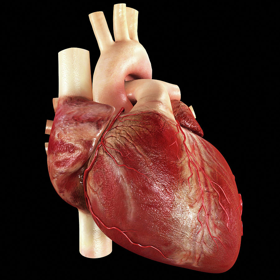
Many of us have a general idea of what the heart looks like, but have you ever seen a real picture of it? The human heart is a muscular organ that is roughly the size of your fist. It is located in the center of your chest, slightly to the left. The heart is divided into four chambers: the right atrium, the right ventricle, the left atrium, and.
Real Human Heart Cliparts.co
The human heart is located in the center of the chest - slightly to the left of the sternum (breastbone). It sits between your lungs and is encased in a double-walled sac called the pericardium.
Human Heart Wallpapers Wallpaper Cave
The heart is composed of smooth muscle. It has four chambers which contract in a specific order, allowing the human heart to pump blood from the body to the lungs and back again with high efficiency. The heart also contains "pacemaker" cells which fire nerve impulses at regular intervals, prompting the heart muscle to contract.
Human Heart Wallpapers Wallpaper Cave
The human heart beats up to 3 billion times over an average lifespan. Learn about the anatomy of the heart and how this muscular organ provides life-giving o.
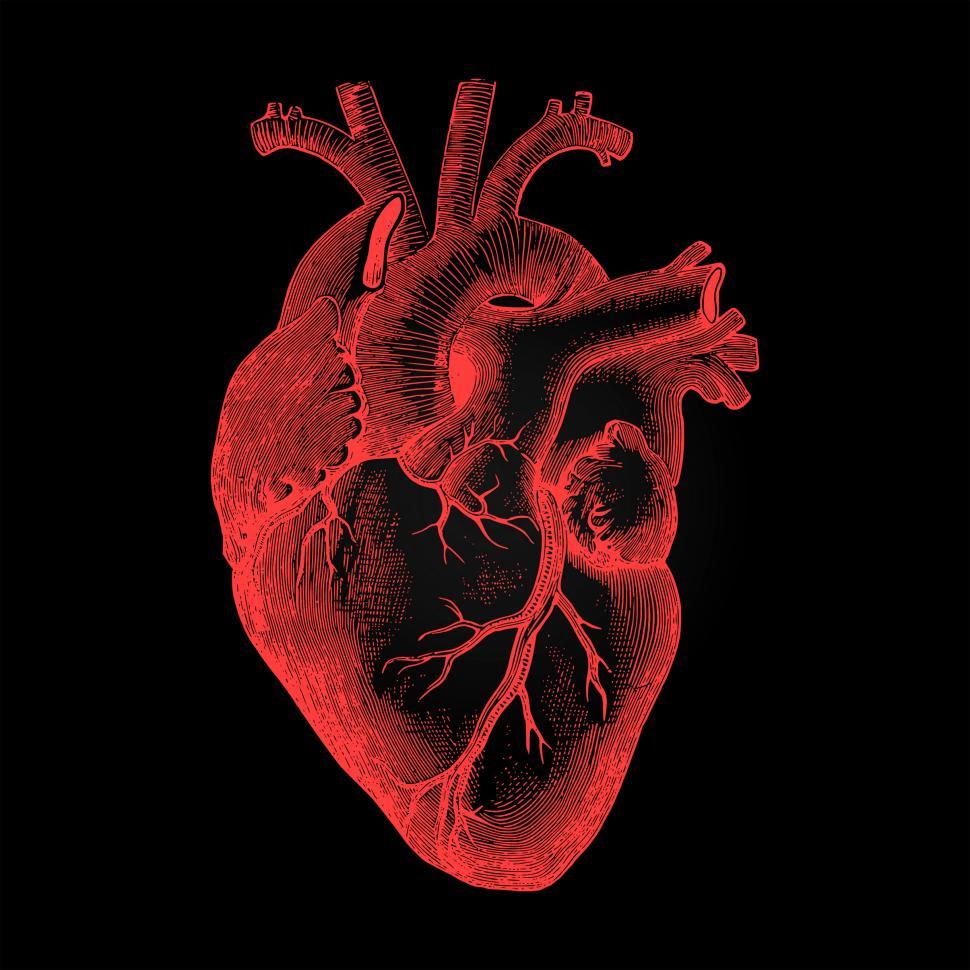
Show me a diagram of the human heart? Here are a bunch!
PLACING THE HEART IN THE CHEST. One of the first rules of human anatomy is that all bodily parts should be described as viewed in the so‐called anatomical position (Anderson and Loukas, 2009).This means that the heart should be described as it is normally positioned within the thorax (Fig. (Fig.1; 1; Cosío et al., 1999; Anderson et al., 2013).It is difficult in the dissecting room, however.